
A Comprehensive Guide to HCG Reference Values Throughout Pregnancy, 48-Hour Growth Standards, and Debunking the Doubling Myth: Interpreting Results After IVF Embryo Transfer, Identifying Ectopic Pregnancy Risks, and Understanding Your HCG Report Scientifically
Read allAfter a positive pregnancy test,Human Chorionic Gonadotropin(HCG) The fluctuation in its value is the metric most closely watched by many pregnant women: What should it be at 4 weeks of pregnancy? If it doesn't double within 48 hours, does that mean the embryo isn't developing well? Could a low value indicate an ectopic pregnancy?
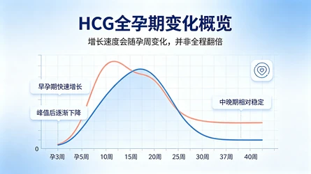
These issues never have a single standard answer. The most core characteristic of HCG is that it rises rapidly in early pregnancy, but there is significant individual variation among normal pregnancies; as gestational weeks increase, the rate of rise gradually slows down, and after reaching its peak, it undergoes a physiological decline.A single value does not have an absolute "normal standard"; it must be interpreted in conjunction with gestational age, dynamic changes, clinical symptoms, and ultrasound findings..
This article compiles HCG reference data for weeks 3–40 of pregnancy, detailing the 48-hour doubling rule, the clinical significance of abnormal values, and natural pregnanciesTest tube babyInterpretation principles after transplantation help you shift from simply comparing numerical values to making evidence-based medical decisions.
Medical tipThis article is for medical education and statistical reference only and does not constitute a basis for individual diagnosis. hCG alone cannot confirm the location of a pregnancy or its final outcome. If you experience sudden severe abdominal pain, shoulder pain, fainting, or heavy vaginal bleeding, seek emergency medical care immediately; do not wait for follow-up tests.
1. Understanding HCG: The Core Signal in Early Pregnancy
HCG, short for human chorionic gonadotropin, is primarily secreted by the syncytiotrophoblast cells of pregnancy tissue. After embryo implantation, trophoblast cells begin to secrete HCG, stimulating the maternal corpus luteum in the ovaries to continue secretingProgesteronemaintains endometrial stability to support early pregnancy; once the placenta matures, it gradually assumes the function of hormone secretion.
1. What HCG Can and Cannot Indicate
HCG only reflects the hormonal secretory activity of gestational trophoblastic cells and cannot be directly equated with embryo quality:
✅Can serve as a biological marker for early pregnancy
✅Dynamic changes can assist in evaluating the trend of pregnancy progression.
❌It is not possible to directly determine whether the embryonic chromosomes are normal.
❌Cannot diagnose fetal structural abnormalities
❌Cannot confirm intrauterine pregnancy or ectopic pregnancy
❌No guarantee of a final live birth outcome
2. Why is HCG undetectable before implantation?
HCG is only gradually introduced into the maternal bloodstream after implantation, when trophoblastic tissue begins to form. In natural pregnancies, implantation typically occurs 6–10 days after ovulation, with significant individual variation. Therefore, if implantation has not yet occurred after fertilization, HCG may be undetectable in both blood and urine tests. This explains why early pregnancy tests can initially yield negative results but turn positive upon retesting a few days later.
3. Differences Between Total HCG and β-HCG
The HCG molecule consists of two subunits, α and β. The α subunit is structurally similar to luteinizing hormone (LH), follicle-stimulating hormone (FSH), and others; the β subunit has higher specificity and serves as the primary target for quantitative clinical pregnancy testing, commonly reported as β-HCG.
Different detection platforms vary in their ability to identify intact hCG and its molecular subtypes.For continuous monitoring, it is recommended to use the same laboratory and the same testing platform., reducing system errors.
4. Blood HCG vs. Urine HCG: Which One to Choose?
Table
Comparison Items | Blood HCG | Urine HCG |
|---|---|---|
Detection Type | Primarily focused on quantitative detection | Primarily qualitative detection |
Result Format | Specific values | Positive / Negative |
Sensitivity | Higher, allowing for earlier detection | Slightly delayed due to threshold limitations |
Core Purpose | Monitoring Dynamic Changes in Early Pregnancy | Preliminary confirmation of pregnancy |
Limitations | The location of the pregnancy cannot be determined independently. | Unable to assess the magnitude of growth |
For the assessment of early pregnancy progression, serum quantitative β-HCG testing is clinically preferred.
II. HCG Reference Range Throughout the Entire Pregnancy: Variation Patterns from Weeks 3 to 40
1. First, let's clarify: How is gestational age calculated?
Clinical gestational age is uniformly calculated based onFirst day of last menstrual period (LMP)Calculated from the last menstrual period, not the date of fertilization. In a standard 28-day menstrual cycle, ovulation occurs approximately 14 days after the last menstrual period; therefore, clinical gestational age is about two weeks longer than the actual time since fertilization:
2 weeks post-fertilization ≈ 4 weeks clinical gestation
3 weeks after fertilization ≈ 5 clinical weeks of pregnancy
4 weeks after fertilization ≈ 6 weeks of clinical pregnancy
For patients with irregular menstrual cycles or delayed ovulation, the gestational age estimated from the last menstrual period may not correspond to the actual stage of pregnancy and requires correction based on ultrasound findings.
2. Reference Table of Serum HCG Levels During Weeks 3–18 of Pregnancy
The following data is derived from the publicly available serum hCG reference intervals provided by MedlinePlus, a service of the U.S. National Library of Medicine. This information is for statistical reference only; reference ranges may vary between laboratories and should not be used directly as diagnostic criteria.
Table
Gestational age (calculated from the last menstrual period) | HCG Reference Range (mIU/mL) |
|---|---|
Not pregnant | <5 |
3 weeks pregnant | 5 – 72 |
4 weeks pregnant | 10 – 708 |
5 weeks pregnant | 217 – 8245 |
6 weeks pregnant | 152 – 32177 |
7 weeks pregnant | 4059 – 153767 |
8 weeks pregnant | 31366 – 149094 |
9 weeks pregnant | 59109 – 135901 |
10 weeks pregnant | 44186 – 170409 |
12 weeks pregnant | 27107 – 201165 |
14 weeks pregnant | 24302 – 93646 |
15 weeks pregnant | 12540 – 69747 |
16 weeks pregnant | 8904 – 55332 |
17 weeks pregnant | 8240 – 51793 |
18 weeks pregnant | 9649 – 55271 |
Note: Reference ranges from different studies may overlap (e.g., the lower limit at 6 weeks of gestation may be lower than that at 5 weeks). This is due to differences in study populations and testing methods, and does not indicate that hCG levels decline with advancing gestational age. Do not interpret this table as a growth curve requiring strict weekly targets.
3. Characteristics of HCG changes during gestational weeks 19–40
In the second and third trimesters, HCG patterns are entirely different from those in early pregnancy: levels gradually decline after peaking and then remain stable. There is no standard for week-by-week increases, and the "doubling" requirement no longer applies.
Table
Gestational Week Stage | Characteristics of HCG Changes | Interpretation Principles |
|---|---|---|
Gestational Weeks 19-24 | In the post-peak decline phase | No longer require 48-hour growth; do not assess development separately |
25-28 weeks of pregnancy | Maintain a relatively stable level | Make a comprehensive assessment based on routine prenatal check-ups |
29-32 weeks pregnant | No rapid growth in the first trimester | Do not use HCG as the core indicator of fetal development |
33-36 weeks of pregnancy | Maintain a relatively stable platform | Interpretation based on clinical indications |
37-40 weeks of gestation | No unified target value | Primarily based on routine obstetric assessment |
4. Why is there such a large variation in HCG levels at the same gestational age?
Individual numerical differences are a normal phenomenon, with the main influencing factors including:
Individual differences in ovulation and implantation timing
Natural differences in secretory activity of trophoblasts
Difference between singleton and multiple pregnancies
Detection of Systematic Differences Between Platforms and Reagents
Margin of error in gestational age estimation
Never treat the upper limit of the reference range as an "ideal target."A single value that is not at the median level does not indicate a pregnancy abnormality.
5. Unit Conversion Explanation
In HCG testing,1 IU/L = 1 mIU/mLThe two values are numerically equivalent, differing only in their unit expressions. If the report uses other units such as ng/mL, do not directly apply this conversion; instead, first verify the specific assay and the corresponding conversion factor.
III. HCG Growth and Peak: It Does Not Need to Double Throughout the Entire Process
HCG does not continue to increase rapidly throughout the entire pregnancy; its overall changes follow a clear physiological pattern: levels begin to rise after implantation → increase rapidly during early pregnancy → peak at approximately 8–10 weeks of gestation → gradually decline → remain stable at low levels during mid- and late pregnancy.
The peak windows vary slightly across different studies (some falling within gestational weeks 9–12), and there is no absolute, universally defined “turning-point gestational week.”
Why doesn't HCG always double?
The rate of HCG increase is determined by the proliferative activity of trophoblast cells, placental development, and hormone metabolism. During early pregnancy, rapid proliferation of trophoblast cells leads to a steep rise in HCG levels. As pregnancy progresses, the placenta gradually assumes the function of the corpus luteum, trophoblast proliferation slows down, and the rate of HCG increase naturally declines; the decrease after reaching peak levels represents normal physiological changes.
The focus of interpretation should gradually shift across different gestational weeks:
Gestational weeks 4–5: Focus on monitoring early growth trends
5–6 weeks pregnant: Use ultrasound to confirm the gestational sac’s location and development
Weeks 6–8 of pregnancy: Based primarily on ultrasound findings such as the fetal pole and fetal heartbeat
Weeks 8–12 of pregnancy: Rapid first-trimester growth is no longer required
Second and Third Trimester: Doubling criteria are completely inapplicable
Cessation of doubling ≠ Embryonic demiseThe key lies in the stage of pregnancy and whether ultrasound provides more direct evidence of development.
IV. The 48-Hour Growth Rule: Debunking the Myth of "Must Double"
The belief that “HCG must double within 48 hours” is the most widespread misconception. Clinical studies have confirmed that HCG growth in normal pregnancies exhibits a wide range of variation and is closely related to initial HCG levels.
1. Classic Studies: The Actual Growth Rate of Normal Pregnancy
The 2004 study by Barnhart et al., published in Obstetrics & Gynecology, included 287 subjects with ultimately confirmed intrauterine viable pregnancies and analyzed 861 serial hCG measurements to establish growth reference ranges for normal pregnancy:
Table
Detection Interval | Median Growth Multiple | Median growth rate | Minimum Growth Reference (1st Percentile) |
|---|---|---|---|
24 hours | 1.50x | +50% | +24% |
48 hours | 2.24 times | +124% | +53% |
96 hours | 5.00x | +400% | No unified minimum value |
The core conclusion of this study:
124% growth in 48 hours isPopulation median, not a benchmark for all normal pregnancies;
The final confirmation of a normal intrauterine pregnancy requires a minimum hCG increase of 53% over 48 hours;
HCG increases approximately linearly on a logarithmic scale, and there is no fixed "doubling rate."
2. Tiered thresholds: The higher the initial HCG, the lower the minimum growth requirement
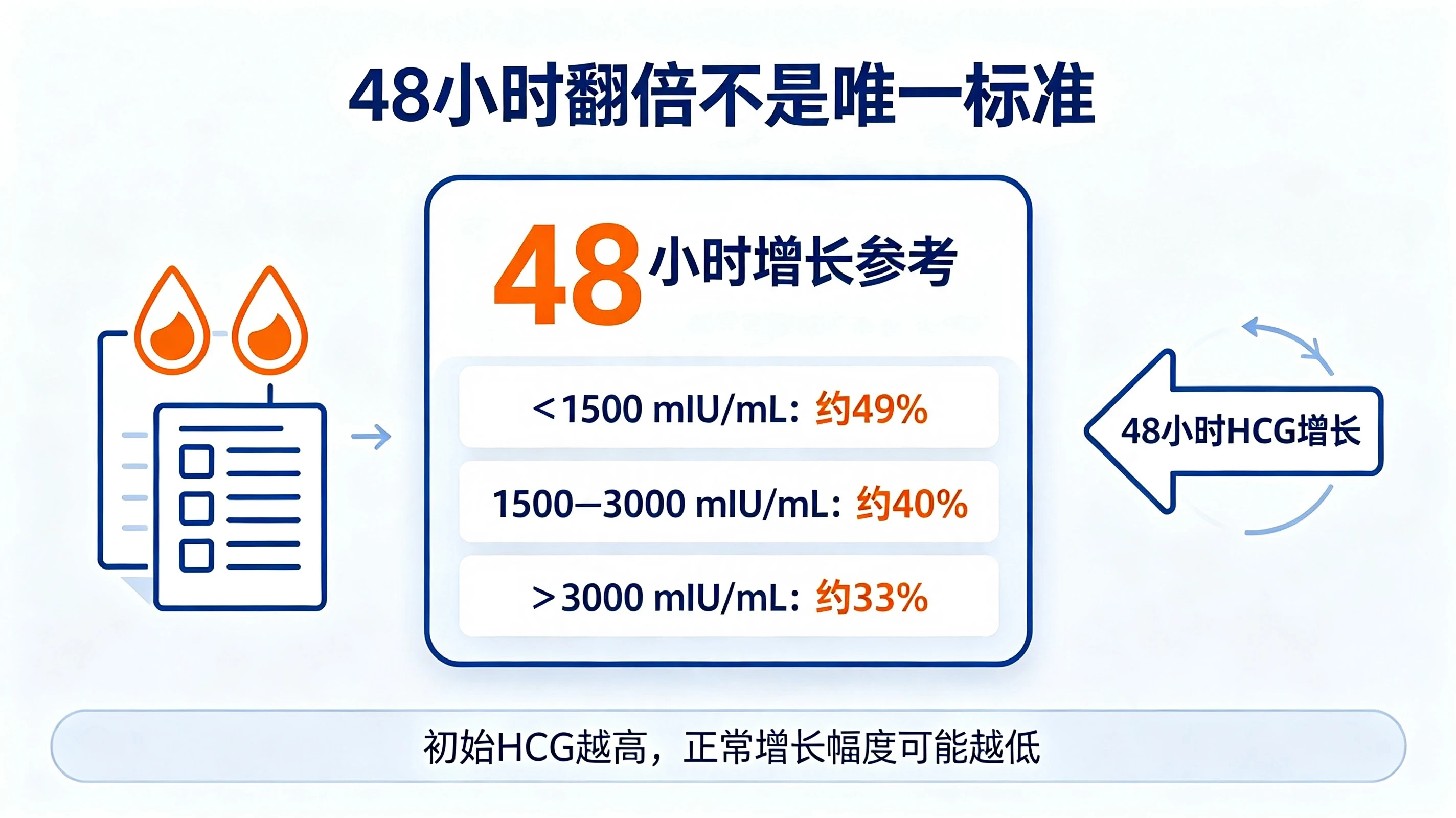
In 2016, the Barnhart team further investigated the minimum rate of increase at different initial hCG levels, including 285 women with first-trimester bleeding or abdominal pain, pregnancy of unknown location, and confirmed intrauterine viable pregnancies. The results showed that the higher the initial hCG level, the lower the minimum increase in normal pregnancies:
Table
Initial serum HCG level | 48-Hour Minimum Growth Reference (1st Percentile) |
|---|---|
<1500 mIU/mL | Approximately 49% |
1500 – 3000 mIU/mL | Approximately 40% |
>3000 mIU/mL | Approximately 33% |
Important note: This data represents the statistical lower limit from a specific study population and is for reference only; it does not constitute clinical diagnostic criteria. It must not be used directly to determine pregnancy location, nor can it serve as a sole basis for decisions regarding termination of pregnancy or treatment of ectopic pregnancy.
3. 35% Growth Line: Reference Boundary for Conservative Follow-up
In 2006, Seeber et al. conducted a study on individuals with unknown pregnancy location and proposed that35% growth in 48 hoursAs a conservative follow-up threshold, the aim is to minimize premature intervention for potentially viable intrauterine pregnancies.
The study also confirmed that the HCG change curves of some ectopic pregnancies are highly similar to those of normal intrauterine pregnancies and spontaneous abortions. Therefore:
An increase of ≥35% in 48 hours cannot definitively prove a normal intrauterine pregnancy.
If the increase within 48 hours is less than 35%, pregnancy failure cannot be directly determined;
Even if HCG levels increase within the reference range, ultrasound is still required to rule out ectopic pregnancy.
35% is a conservative threshold for cautious follow-up, not a passing mark for normal pregnancy.
4. 48-hour growth rate calculation formula
\(\text{48-Hour Growth Rate} = \frac{\text{Second HCG Value} - \text{First HCG Value}}{\text{First HCG Value}} \times 100\%\)
Example:
First HCG level of 100 mIU/mL, rising to 160 mIU/mL after 48 hours: a growth rate of 60%. Although it did not double, this does not indicate an abnormal pregnancy.
First HCG level: 2000 mIU/mL; after 48 hours: 2800 mIU/mL (40% increase). Interpretation requires integration of the initial level, gestational age, and ultrasound findings.
If the interval between two blood draws is not 48 hours, do not directly apply the above percentages; a doctor must assess based on the actual interval.
V. How to Interpret Abnormal HCG Levels? Clinical Significance of High, Low, or Slowly Rising Levels
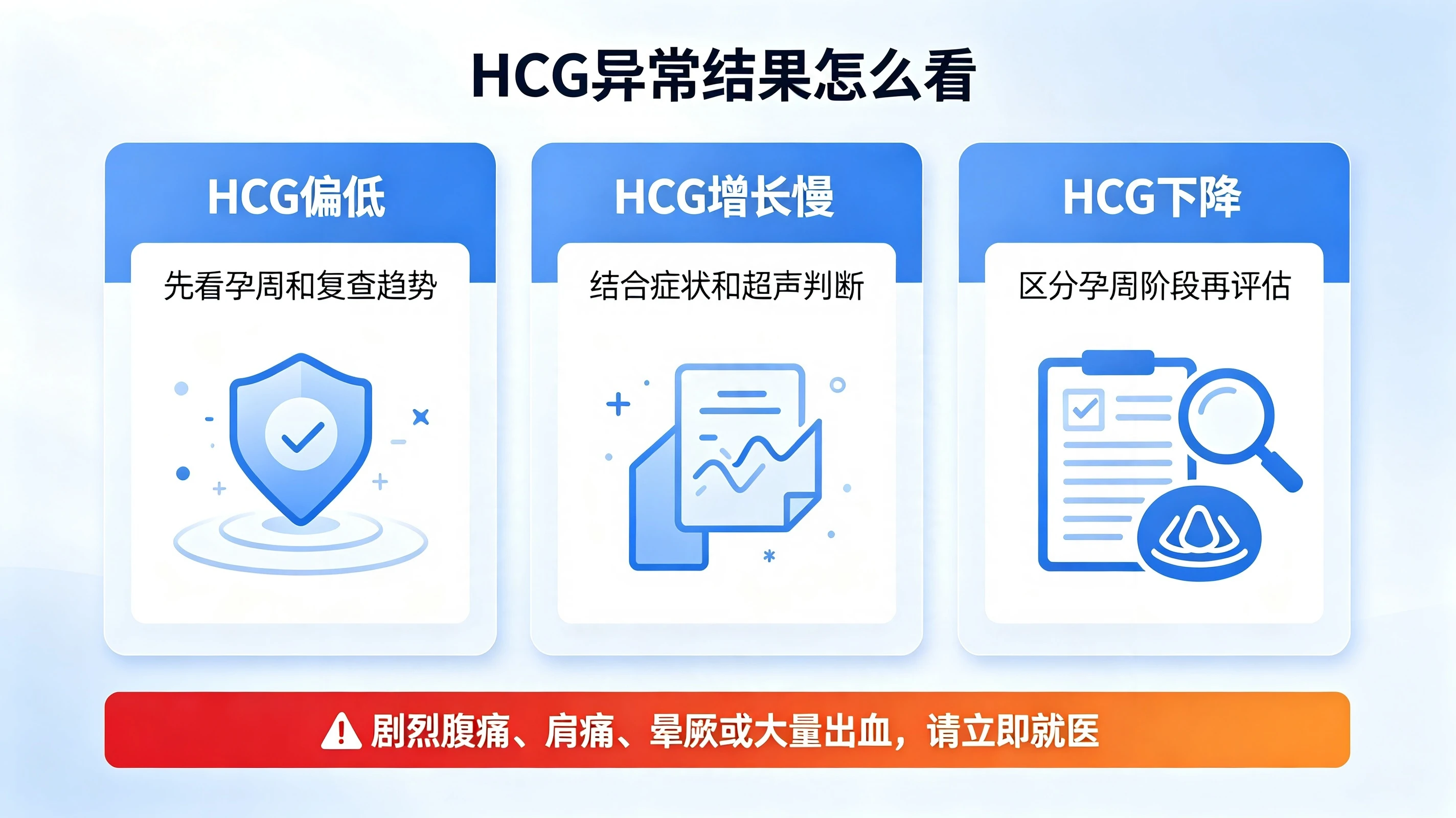
If an abnormal result is obtained, it is not recommended to draw conclusions independently. Clinicians will first verify the gestational age and testing conditions, then make a comprehensive assessment by combining dynamic trends with ultrasound findings.
1. HCG level below the reference range for gestational age
Possible causes:
Ovulation/implantation occurred later than estimated, resulting in an actual gestational age that is less than the gestational age calculated from the last menstrual period.
Trophoblast-secreted activity is slightly low
Biochemical pregnancy or early pregnancy loss
Ectopic pregnancy
Recommended ActionsA single low value is insufficient to determine the outcome. A follow-up test should be conducted approximately 48 hours later under medical guidance, with an ultrasound monitoring plan established simultaneously, focusing on whether the values continue to rise.
2. Slow HCG increase
Possible causes:
Normal intrauterine pregnancy, but growth is at the low end of the population range.
Early pregnancy loss in progress
Ectopic pregnancy
Deviation in gestational age or blood draw timing
Key ReminderSlow growth is not a definitive diagnostic criterion for ectopic pregnancy; further evaluation combining ultrasound findings and clinical manifestations is required.
3. HCG remains consistently flat
If HCG levels show no significant change in two or more consecutive tests during early pregnancy, heightened vigilance is required, as this may indicate abnormal pregnancy development and can also be seen in ectopic pregnancy. In cases where the location of the pregnancy is unknown, the risk of ectopic pregnancy cannot be ruled out solely because HCG levels have not decreased.
Recommended ActionsSeek medical attention as soon as possible, follow your doctor’s instructions for follow-up visits and complete ultrasound evaluations. Do not extend the observation interval on your own.
4. Decrease in HCG
The nature must be differentiated according to gestational age:
Early pregnancy (pregnancy outcome not yet confirmed): A continuous decline often suggests cessation of embryonic development. The location of the pregnancy must be determined, and follow-up should continue until hCG becomes undetectable or a clinically safe endpoint is reached.
After peaking at 8–10 weeks of pregnancy, a gradual decline is a normal physiological process.
5. Significant elevation of HCG
Possible causes:
Actual gestational age is greater than the estimated gestational age.
Twin or multiple pregnancy
Gestational Trophoblastic Disease, such as Hydatidiform Mole
Other rare conditions or assay interferences that cause hCG secretion
Key ReminderElevated HCG levels do not necessarily indicate good embryo quality and cannot confirm a twin pregnancy. Significantly high levels require ultrasound confirmation of gestational age, fetal number, and the morphology of the pregnancy tissue.
VI. HCG and Progesterone: Not Interchangeable, Nor Equivalent
HCG reflects the secretory activity of trophoblast cells, while progesterone indicates the pregnancy-supporting function of the corpus luteum or placenta; these two are independent markers and cannot be used to infer one another.
High progesterone ≠ Embryo is definitely normalHigh progesterone levels cannot guarantee embryonic survival, nor can they exclude ectopic pregnancy.
Low progesterone ≠ Guaranteed miscarriageProgesterone levels are influenced by multiple factors, including the stage of pregnancy, corpus luteum function, and medication supplementation; in patients undergoing assisted reproductive technology who receive luteal phase support medications, interpretation of serum progesterone levels should be considered in conjunction with their specific medication regimen.
It is not recommended to use a single progesterone cutoff value to predict outcomes.Progesterone cannot reliably distinguish between intrauterine viable pregnancies and ectopic pregnancies; single values such as 25 ng/mL or 15 ng/mL should not be used directly to define "normal" versus "abnormal."
VII. From Blood Draw to Ultrasound: When Should Imaging Tests Be Done?
Blood tests can only reflect hormonal changes, whereas ultrasound is the direct method for confirming the location of pregnancy and observing embryonic development; the integration of these two approaches is central to decision-making in early pregnancy.
1. What is the HCG decision region?
The hCG discriminatory zone refers to the serum hCG level at which an intrauterine gestational sac is likely to be visualized on transvaginal ultrasound. The traditional reference range is 1,500–2,000 mIU/mL; however, to avoid misclassifying normal intrauterine pregnancies as abnormal, the American College of Obstetricians and Gynecologists (ACOG) recommends using a more conservative threshold, with values up to 3,500 mIU/mL being considered acceptable.
HCG < 1500 mIU/mL: It is normal for the gestational sac not to be visible on ultrasound at this level.
HCG 1500–3500 mIU/mL: Requires comprehensive evaluation in conjunction with ultrasound equipment, gestational age, and clinical manifestations;
When hCG levels exceed 3500 mIU/mL without a clearly identified intrauterine gestational sac, further evaluation for ectopic pregnancy is required.
NoteEven if the HCG level exceeds 3500 mIU/mL without visualization of a gestational sac, ectopic pregnancy cannot be definitively diagnosed based on this finding alone; furthermore, it is imperative not to intervene in a potentially viable intrauterine pregnancy when evidence is insufficient.
2. Key ultrasound observation points at different gestational ages
Table
Approximate Gestational Age | Key Points of Ultrasound Observation | Clinical Significance |
|---|---|---|
4-5 weeks pregnant | Is the intrauterine gestational sac visible? | Preliminary confirmation of pregnancy location |
5-6 weeks pregnant | Yolk sac, gestational sac, with the fetal pole partially visible | Assess early developmental status |
6-7 weeks pregnant | Germinal Length and Primitive Heart Tube Pulsation | Confirm embryonic viability |
7-8 weeks pregnant | Germinal growth and cardiac activity | Further verify development |
After 8 weeks of pregnancy | Embryonic growth, fetal heart rate, and other structural indicators | Primarily image-based follow-up |
The timing of ovulation and implantation, ultrasound equipment, and operator experience all affect the observed results; there is no absolute, universally standardized timeline.
3. After seeing the fetal heartbeat, is it still necessary to check HCG levels?
After an ultrasound confirms an intrauterine pregnancy with a visible fetal heartbeat,Routine monitoring of HCG doubling is not requiredSubsequent management should primarily consist of ultrasound follow-up and routine prenatal check-ups.
If special circumstances such as bleeding, abdominal pain, or uncertain pregnancy location are present, the doctor may order HCG tests as needed; however, repeated blood draws to pursue a "perfect curve" without clinical indications are not recommended.
8. Special Considerations for Interpreting HCG Levels After IVF Embryo Transfer
IVF embryo transfer has a clearly defined transfer date, which reduces errors in estimating gestational age; however, hCG levels still cannot directly predict the final outcome.
1. Gestational Age Conversion After Embryo Transfer
Clinical gestational age is still calculated based on the last menstrual period (LMP), with the conversion formula as follows:
Day 5 Blastocyst Transfer: Transfer day = 2 weeks and 5 days pregnant, gestational age in days = days after transfer + 19
Day 3 Embryo Transfer: Day of transfer = 2 weeks and 3 days pregnant; Gestational age in days = Days post-transfer + 17
Table
Transplant Type | Days Post-Transplant | Corresponding clinical gestational age |
|---|---|---|
Day 5 Blastocyst | 9dp5dt | 4 weeks 0 days pregnant |
Day 5 Blastocyst | 10dp5dt | 4 weeks and 1 day pregnant |
Day 5 Blastocyst | 12dp5dt | 4 weeks and 3 days pregnant |
Day 3 Embryo | 12dp3dt | 4 weeks and 1 day pregnant |
Day 3 Embryo | 14dp3dt | 4 weeks and 3 days pregnant |
2. There is no standardized "normal value" for HCG in the early post-transplant period
There is no standard value applicable to all patients; influencing factors include:
Developmental stage of the transferred embryo (Day 3 embryo / Day 5 blastocyst)
Actual embryo implantation time
Number of Embryos Transplanted
Discrepancies Between the Detection Platform and the Laboratory
Whether exogenous hCG is used during the ovarian stimulation phase and its metabolic time
Never treat integer values such as 100 mIU/mL or 150 mIU/mL as universal safety thresholds.
3. Big Data Reference: HCG and Live Birth Prediction After Frozen-Thawed Blastocyst Transfer
A 2025 single-center retrospective study in F&S Reports analyzed 18,491 frozen-thawed blastocyst transfer cycles and determined the predictive cutoff for human chorionic gonadotropin (HCG) levels at 7–11 days post-transfer for live birth:
Table
Days Post-Transplant | Live Birth Prediction Cut-off (mIU/mL) |
|---|---|
Day 7 | 31.05 |
Day 8 | 46.60 |
Day 9 | 72.05 |
Day 10 | 95.40 |
Day 11 | 159.90 |
Important note: This value is a statistical predictive cutoff, not a diagnostic criterion for pregnancy viability. The study is based on single-center retrospective data and does not apply to day-3 embryo transfer. Population-level predicted probabilities cannot be directly equated with individual outcomes and should not be used as a basis for discontinuing treatment.
4. Live birth is still possible even with low initial HCG levels
A low initial hCG level does not necessarily indicate pregnancy failure; it should be evaluated in conjunction with the number of days post-transfer, embryo type, subsequent 48-hour hCG rise, history of exogenous medication use, and ultrasound findings. Predicting the final outcome based solely on the first hCG value is not recommended.
5. Singleton or twin pregnancy cannot be determined solely based on HCG levels
In multiple pregnancies, average hCG levels are higher, but the numerical ranges for single and multiple pregnancies overlap significantly. A high hCG level does not indicate a twin pregnancy, nor does a low level guarantee a singleton; the final number of fetuses is determined by ultrasound examination.
9. Be Wary of Ectopic Pregnancy: HCG Alone Cannot Rule Out the Risk
Ectopic pregnancy (most commonly tubal pregnancy) is the most dangerous complication of early pregnancy and requires emphasis:The HCG profile in ectopic pregnancy is nonspecific.Some may exhibit slow growth, remain stable, or even show growth patterns similar to those of a normal intrauterine pregnancy, while others may decline.
Seek immediate emergency care if these symptoms appear
The following symptoms may indicate intra-abdominal bleeding, which is a medical emergency. Do not wait for a follow-up appointment:
Severe pain in one lower quadrant of the abdomen, progressively worsening.
Sudden severe abdominal pain
Radiating pain in the shoulder, accompanied by abdominal pain and dizziness
Obvious dizziness, fatigue, and syncope
Heavy vaginal bleeding
2. The absence of symptoms does not rule out ectopic pregnancy
Some cases of ectopic pregnancy may present with no obvious abdominal pain or bleeding in the early stages. If intrauterine pregnancy is not confirmed by ultrasound, it is essential to follow medical advice for continuous monitoring through serial hCG measurements and transvaginal ultrasonography; vigilance should not be relaxed merely because symptoms are absent.
3. Normal HCG growth does not rule out ectopic pregnancy
HCG levels only reflect hormone secretion and cannot indicate the implantation site of the embryo. Even if the increase falls within the normal reference range for pregnancy, ectopic pregnancy cannot be 100% ruled out.
10. Standard Decision-Making Process After Receiving the HCG Report
Step 1: Verify basic information
Confirm the testing time, HCG values and units, testing institution, last menstrual period or embryo transfer date, whether any HCG-containing medications were used, and clarify the actual interval between the two blood draws.
Step 2: Determine whether a re-examination is required
First confirmation of pregnancy, assessment of progression required: Follow doctor's orders to recheck approximately 48 hours later.
With HCG levels continuing to rise and ultrasound confirming an intrauterine pregnancy, the doctor will determine whether further blood tests are needed.
Step 3: Calculate the growth rate and interpret it by layer
Calculate the percentage change between two HCG measurements and interpret it in conjunction with the initial level: a lower initial HCG predicts faster growth; a higher initial HCG corresponds to a lower minimum growth threshold; after 8 weeks of gestation, early pregnancy growth standards no longer apply.
Step 4: Assess the timing of ultrasound examination
Schedule a transvaginal ultrasound promptly once the gestational age is appropriate or hCG levels enter the discriminatory zone. Do not delay necessary ultrasounds solely because hCG has not reached a specific threshold, and do not diagnose ectopic pregnancy based on a single scan without visualization of an intrauterine gestational sac.
Step 5: Clarify the key points for subsequent follow-up
Table
Current situation | Core basis for judgment | Next Steps |
|---|---|---|
First positive pregnancy test | HCG + Gestational Age Calculation | Follow-up appointments as directed by your doctor |
HCG levels are low but steadily rising | Dynamic Trend | Continue to standardize follow-up |
Slow HCG increase | HCG trends + symptoms + ultrasound | Seek timely medical evaluation |
HCG remains flat or decreases | Pregnancy Location + Clinical Manifestations | Seek medical attention as soon as possible for further examinations. |
Elevated HCG levels with no gestational sac visible on ultrasound | Ultrasound + Continuous HCG | Investigation of Pregnancy of Unknown Location and Ectopic Pregnancy |
Intrauterine fetal viability confirmed. | Ultrasound Developmental Indicators | Follow up according to the prenatal checkup schedule |
Sudden severe abdominal pain / Syncope | Clinical Symptoms | Immediate Emergency |
Recommendation: Establish personal follow-up records
Record the following information at each check-up to help your doctor make a better assessment: date and time of blood draw, HCG value and unit, actual interval duration, growth rate, estimated gestational age, symptoms (such as abdominal pain, bleeding, or dizziness), and ultrasound results (gestational sac, yolk sac, fetal pole, and fetal heartbeat).
XI. Pitfall Guide: 8 Common Misconceptions in HCG Interpretation
❌ MisconceptionThe higher the HCG, the better the embryo quality.✅Correct answer: HCG only reflects trophoblast activity and has no direct correlation with embryonic chromosomal or structural health.
❌ MisconceptionIf hCG levels do not double within 48 hours, the pregnancy cannot be saved.✅Correct answer: Doubling represents the median among the population; in normal pregnancies, the minimum growth rate can be significantly below 100%, with considerable individual variation.
❌ Misconception: An increase of more than 35% within 48 hours can rule out ectopic pregnancy.✅Correct answer: The HCG growth curve in partial ectopic pregnancy is similar to that of a normal pregnancy, so it cannot be ruled out based on the growth pattern alone.
❌ MisconceptionA low HCG level relative to the gestational age indicates poor embryonic development.✅Correct answer: Late ovulation and implantation leading to gestational age discrepancy is the most common cause, which requires assessment based on trends.
❌ MisconceptionThe higher the HCG level, the greater the probability of a twin pregnancy.✅Correct answer: The average level is higher in multiple pregnancies, but the numerical ranges overlap significantly; definitive diagnosis can only be made via ultrasound.
❌ MisconceptionHigh progesterone levels do not guarantee normal embryonic development.✅Correct answer: Progesterone alone cannot confirm embryonic viability, nor can it rule out ectopic pregnancy.
❌ MisconceptionAfter the fetal heartbeat is detected, HCG must continue to double✅Correct answer: HCG growth rate slows down with gestational age, peaks and then declines; after fetal heart activity is detected, ultrasound follow-up becomes the primary monitoring method.
❌ MisconceptionA decline in HCG does not necessarily indicate fetal arrest.✅Correct answer: A sustained decline in early pregnancy warrants caution for pregnancy loss; a decline after the peak is part of normal physiological changes.
Summary: Don't chase impressive numbers; focus on identifying the correct decision points.
HCG is a highly valuable monitoring indicator in early pregnancy, but it has never been a "report card on embryo quality," let alone a "verdict on pregnancy outcomes."
Interpreting HCG is fundamentally not about comparing absolute values with others, but rather about assessing whether the gestational age calculation is accurate, whether a single measurement aligns with the current stage of pregnancy, whether dynamic changes warrant further evaluation, whether the location of the pregnancy has been confirmed by ultrasound, and whether there are risks of acute complications.
For patients with an unknown location of pregnancy, serial hCG measurements are a useful tool for risk identification but cannot replace ultrasound. For those with a confirmed intrauterine viable pregnancy, monitoring hCG doubling is far less meaningful than standard ultrasound examinations and routine prenatal care.
What matters most in early pregnancy is not achieving satisfactory results with every blood test, but completing the appropriate examinations at the right time—neither missing risks such as ectopic pregnancy nor disrupting a potentially normal pregnancy through premature or excessive interpretation.
Frequently Asked Questions
The content on this website is general educational material and compiled public information about IVF. It is provided for reference only, does not constitute a basis for diagnosis or medical decision-making, and does not constitute any medical advice or treatment recommendation. For policy and technical requirements, please refer to official publications issued by the relevant authorities and institutions. If you find any content errors, numerical inconsistencies, or outdated information, please contact us so we can verify and update it.